top of page
מאמרים
ריכזתי כאן קישורים למאמרים אקדמיים בנושאים שונים שפרסמתי בכתבי עת מובילים לאורך השנים. המאמרים עסקו בשאלות רלוונטיות המאתגרות את הגישות והשיטות הקיימות, בעיקר לניתוח החלפת מפרק ירך וניתוח החלפת מפרק ברך אך ישנם מאמרים גם בתחומים נוספים. במידה ואתם מעוניינים לקבל מידע נוסף או לקרוא את המאמרים המלאים לחצו על המאמר הרלוונטי לכם אותו תרצו לקרוא ותקבלו את תמצית המאמר וקישור למאמר המלא. קריאה מהנה.
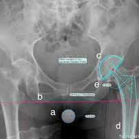
Radiographic templating of total hip art
Abstract |Purpose: The purpose of this study was to evaluate the use of pre-operative digital templating to minimize complications including limb length discrepancy (LLD), intraoperative fractures and early dislocations in patients with intracapsular femoral neck fractures.
Methods: We retrospectively compared 23 patients undergoing total hip arthroplasty (THA) for intracapsular femoral fractures with pre-operative digital templating and 48 patients without templating.
Results: The mean post-operative LLD was significantly lower in patients who had pre-operative templating than in the control group (6.7 vs. 11.5 mm, p = 0.023). Only three patients (13 %) with templating had LLD greater than 1.5 cm, compared to the 15 patients (31 %) without templating (p = 0.17). In eight cases the final femoral stem size matched the templated size, while 19 patients were within two size increments. Complications included one dislocation and one intra-operative fracture in the control group.
Conclusion: The present study demonstrated that careful pre-operative planning may reduce LLD in patients undergoing THA due to intracapsular hip fractures.
Keywords: Femoral neck fracture; Leg length discrepancy; Pre-operative planning; Templating; Total hip arthroplasty.

Importance of the dome and posterior wal
Background: Characterizing the distribution of bone density in the acetabulum is of importance in better understanding and guiding treatment for both osteoarthritis and trauma of the hip joint. This study aims to develop a highly automated method to quantify the pattern of subchondral bone density in the acetabulum using clinically identifiable regions.
Methods: Subchondral acetabular bone density distribution maps were created bilaterally from 30 non-pathologic pelvic CT scans. The density maps were aligned orthogonal to the acetabular rim plane and divided into twelve zones. Average bone density was calculated in each of these zones and compared to investigate differences between regions within each acetabulum and between right and left sides in a given patient.
Findings: In all patients, the zones corresponding to the dome and posterior wall of the acetabulum demonstrated significantly higher average bone

Proximal Femoral Shortening After Cephalomedullary Nail Insertion for Intertrochanteric Fractures
Abstract | Objective: To assess the incidence of proximal femoral shortening (PFS) and its effect on the patient outcomes when intertrochanteric fractures were treated with a cephalomedullary nail (CMN).
Design: Retrospective cohort study.
Settings: Level II trauma center.
Patients: Forty-eight consecutive patients with OTA/AO 31-A intertrochanteric fractures.
Intervention: All patients were treated with a Gamma3 CMN (Stryker, Kalamazoo, MI).
Methods: PFS was assessed for abductor lever arm (x vector), femoral height (y vector), and overall shortening (z vector) on anteroposterior radiographs. Fixation success and retained ambulatory capacity were noted.
Results: Shortening of >5 mm of the x, y, and z vectors was evident in 18, 20, and 29 patients, respectively. Shortening of >10 mm of the x, y, and z vectors was measured in 5, 6, and 8 patients, respectively. Mean shortening of the x, y, and z vectors was 4.5, 5.5, and 7 mm, respectively. Greater PFS was found to be associated with fixation failure and inability to retain ambulatory capacity, independently (P ≤ 0.05 and P ≤ 0.025, respectively). Of note, an unstable fracture pattern was not found to be associated with greater PFS.
Conclusions: PFS is a common phenomenon after CMN of intertrochanteric fractures with a Gamma CMN. In addition, greater PFS seems to be associated with fixation failure and inability to retain ambulatory capacity postoperatively.
Level of evidence: Therapeutic Level IV. See Instructions for Authors for a complete description of levels of evidence.
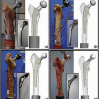
Patient-specific finite element analysis of femurs with cemented hip implants
Abstract | Background: Over 1.6 million hip replacements are performed annually in Organisation for Economic Cooperation and Development countries, half of which involve cemented implants. Quantitative computer tomography based finite element methods may be used to assess the change in strain field in a femur following such a hip replacement, and thus determine a patient-specific optimal implant. A combined experimental-computational study on fresh frozen human femurs with different cemented implants is documented, aimed at verifying and validating the methods.
Methods: Ex-vivo experiments on four fresh-frozen human femurs were conducted. Femurs were scanned, fractured in a stance position loading, and thereafter implanted with four different prostheses. All femurs were reloaded in stance positions at three different inclination angles while recording strains on bones' and prosthesis' surfaces. High-order FE models of the intact and implanted femurs were generated based on the computer tomography scans and X-ray radiographs. The models were virtually loaded mimicking the experimental conditions and FE results were compared to experimental observations.
Findings: Strains predicted by finite element analyses in all four femurs were in excellent correlation with experimental observations FE = 1.01 × EXP - 0.07,R2 = 0.976, independent of implant's type, loading angle and fracture location.
Interpretation: Computer tomography based finite element models can reliably determine strains on femur surface and on inserted implants at the contact with the cement. This allows to investigate suitable norms to rank implants for a patient-specific femur so to minimize changes in strain patterns in the operated femur.
Keywords: Femur; P-FEMs; Total hip arthroplasty.
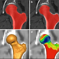
Subchondral bone density distribution in
Abstract | Objective: This study aims to quantitatively characterize the distribution of subchondral bone density across the human femoral head using a computed tomography derived measurement of bone density and a common reference coordinate system.
Materials and methods: Femoral head surfaces were created bilaterally for 30 patients (14 males, 16 females, mean age 67.2 years) through semi-automatic segmentation of reconstructed CT data and used to map bone density, by shrinking them into the subchondral bone and averaging the greyscale values (linearly related to bone density) within 5 mm of the articular surface. Density maps were then oriented with the center of the head at the origin, the femoral mechanical axis (FMA) aligned with the vertical, and the posterior condylar axis (PCA) aligned with the horizontal. Twelve regions were created by dividing the density maps into three concentric rings at increments of 30° from the horizontal, then splitting into four quadrants along the anterior-posterior and medial-lateral axes. Mean values for each region were compared using repeated measures ANOVA and a Bonferroni post hoc test, and side-to-side correlations were analyzed using a Pearson's correlation.
Results: The regions representing the medial side of the femoral head's superior portion were found to have significantly higher densities compared to other regions (p < 0.05). Significant side-to-side correlations were found for all regions (r(2) = 0.81 to r(2) = 0.16), with strong correlations for the highest density regions. Side-to-side differences in measured bone density were seen for two regions in the anterio-lateral portion of the femoral head (p < 0.05).
Conclusions: The high correlation found between the left and right sides indicates that this tool may be useful for understanding 'normal' density patterns in hips affected by unilateral pathologies such as avascular necrosis, fracture, developmental dysplasia of the hip, Perthes disease, and slipped capital femoral head epiphysis.
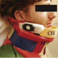
A new external upper airway opening device combined with a cervical collar
Abstract: Airway problems are the main cause of mortality in otherwise survivable trauma injuries. We developed a novel external airway protector in combination with a cervical collar. The new device simultaneously opens the airway and protects the cervical spine.
Materials and methods: The device called the 'Lubo Collar' has a chin holder that can be attached to a gliding knob on the collar. When the knob is pushed forward, the mandible moves forward, thus imitating the jaw thrust manoeuvre and opens the airway. In order to study the safety and efficacy of this new device, a two-phase clinical trial was conducted. In the safety phase 20 patients were evaluated for adverse reactions immediately, 2h and 24h following application of the device. The efficacy phase evaluated the ability of the device to open and maintain an airway in anaesthetised patients. In this phase, 10 patients who had undergone orthopaedic surgery under general anaesthesia were included. Seven patients had blocked airways following anaesthesia induction. The gliding knob attached to the mandible arc was pushed 1cm forward to open their airways.
Results: No adverse events were recorded. In the seven patients with blocked airways, the external airway/collar device opened and maintained patent airways.
Conclusion: The new external non-invasive airway device (Lubo Collar) is safe and effective in opening and maintaining an open airway in an unconscious anaesthetised patient with a blocked airway. These preliminary results may encourage assessment in the field.

Acetabular orientation variability and symmetry based on CT scans of adults
Abstract |Purpose: Understanding acetabular orientation is important in many orthopaedic procedures. Acetabular orientation, usually described by anteversion and abduction angles, has uncertain measurement variability in adult patients. The goals of this study are threefold: (1) to describe a new method for computing patient-specific abduction/anteversion angles from a single CT study based on the identification of anatomical landmarks and acetabular rim points; (2) to quantify the inaccuracies associated with landmark selection in computing the acetabular angles; and (3) to quantify the variability and symmetry of acetabular orientation.
Methods: A total of 25 CT studies from adult patients scanned for non-orthopaedic indications were retrospectively reviewed. The patients were randomly selected from the hospital’s database. Inclusion criteria were adults 20–65 years of age. Acetabular landmark coordinates were identified by expert observers and tabulated in a spreadsheet. Two sets of calculations were done using the data: (1) computation of the abduction and anteversion for each patient, and (2) evaluation of the variability of measurements in the same individual by the same surgeon. The results were tabulated and summary statistics computed.
Results: This retrospective study showed that acetabular abduction and anteversion angles averaged 54° and 17°, respectively, in adults. A clinically significant intra-patient variability of >20° was found. We also found that the right and left side rim plane orientation were significantly correlated, but were not always symmetric.
Conclusion: A new method of computing patient-specific abduction and anteversion angles from a CT study of the anterior pelvic plane and the left and right acetabular rim planes was reliable and accurate. We found that the acetabular rim plane can be reliably and accurately computed from identified points on the rim. The novelty of this work is that angular measurements are performed between planes on a 3-D model rather than lines on 2-D projections, as was done in the past.

Early diagnosis of occult hip fractures: MRI versus CT scan
Objective: We compared Computerised Tomography (CT) and Magnetic Resonance Imaging (MRI) in diagnosis of a painful hip in elderly patients after trauma. We report on accuracy, efficiency and benefits.
Design: We assessed 13 patients, average age 73 years, after fall with plain X-rays showing no evidence of fracture. There were two groups: Group A (six patients) underwent CT and MRI; Group B underwent MRI only.
Results: In Group A where all of the six patients underwent CT and MRI, four of the CT images resulted in misdiagnosis due to inaccuracy. In Group B where all the seven patients underwent only MRI, all the results were accurate and enabled a precise and fast diagnosis.
Conclusions: MRI was found to be a more accurate modality than CT scan for obtaining early diagnosis of occult hip fractures. These results point out the advantage of immediate MRI imaging in patients with occult hip fracture enabling a more effective treatment, a shorter hospitalisation period entailing decreased medical costs.
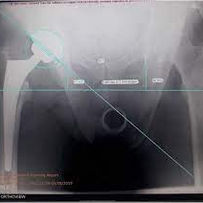
Acetabular Cup Orientation and Postoperative Leg Length Discrepancy in Patients Undergoing Elective Total Hip Arthroplasty via a Direct Anterior and Anterolateral Approaches
Abstract |Background: Total hip arthroplasty (THA) is considered a successful surgical procedure. It can be performed by several surgical approaches. Although the posterior and anterolateral approaches are the most common, there has been increased interest in the direct anterior approach. The goal of the present study is to compare postoperative leg length discrepancy and acetabular cup orientation among patients who underwent total hip arthroplasty through a direct anterior (DAA) and anterolateral (ALA) approaches.
Methods: The study included 172 patients undergoing an elective THA by a single surgeon at our institution within the study period. Ninety-eight arthroplasties were performed through the ALA and 74 arthroplasties through the DAA. Preoperative planning was performed for all patients. Assessment of the two groups included the following postoperative parameters: abduction angle, cup anteversion angle and leg length discrepancy (LLD). Additional analysis was done to evaluate component positioning by comparing deviation from the Lewinnek zone of safety in both approaches.
Results: For the DAA the absolute LLD was 11 mm, ranging from -6 mm to 5 mm. For the ALA, the absolute LLD was 36 mm, ranging from -22 mm to 14 mm. None of the DAA patients had an absolute LLD greater than 6 mm. Comparatively, 7.4% of the ALA group exceeded 6 mm of LLD in addition to 2.1% with LLD greater than 10 mm. 15% of the ALA group resided out of the Lewinnek abduction zone compared to 3% of the DAA group (P = 0.016). 17% of the ALA group were out of the Lewinnek anteversion zone as opposed to 8% of the DAA group (P = 0.094).
Conclusion: Our study demonstrates good component positioning outcomes and LLD values in patients following THA through the DAA compared to the ALA.

Acetabular orientation: anatomical and functional measurement
Abstract | Purpose: Acetabular orientation is important to consider in hip joint pathology and treatment. This study aims to describe the functional orientation of the acetabulum as a representative measure of force transmitted through the hip joint generated from bone density mapping and compare it to landmark-based anatomical orientation measures.
Methods: CT scans of 38 non-pathologic individuals were analyzed. Functional orientation was computed as the density-weighted average of the acetabular surface normals based on surface density maps. Two anatomical measures were also used to describe the orientation of each acetabulum: the normal to the acetabular rim plane and the abduction angle based on AP pelvic "Radiographs" generated from the CT data.
Results: The average functional and anatomic abduction and anteversion angles ranged from 32°-58° and 22°-31°, respectively, with significant side-to-side correlation in individual patients for the majority of measures. Functional acetabular orientation was weakly correlated only with the rim plane measure. Native acetabular abduction in the 3D anatomic and functional methods was significantly shallower than the 2D "Radiographic" measure. The vector generated to describe functional acetabular orientation was found to be more vertically and posteriorly oriented than the anatomic measures.
Conclusions: Functional acetabular orientation, reflecting the calculated directionality of the subchondral bone density, yields a more posterior and vertical measure of acetabular orientation as compared to the direction of load transmission suggested by the anatomic methods.

Abstract Background: Femoral neck geometry directly affects load transmission through the hip. Orientations may be described anatomically or using functional definitions that consider load transmission. Questions/purposes: This study introduces and applies a new method for characterizing functional femoral orientation based on the distribution of subchondral bone density in the femoral head and compares it with orientation measures generated via established anatomic landmark-based methods. Both orientation methods then are used to characterize side-to-side symmetry of orientation and differences between men and women within the population. Patients and methods: A retrospective review of CT imaging data from 28 patients was performed. Anatomic orientation was determined using established two-dimensional and three-dimensional landmarking methods. Subchondral bone density maps were generated and used to define a density-weighted surface normal vector. Orientation angles generated by the three methods were compared, with side-to-side symmetry and differences between genders also investigated. Results: The three methods measured substantially different angles for anteversion and neck-shaft angle. Weak correlations were found between anatomic and functional orientation measures for neck-shaft angle only. Conclusions: Neck-shaft angles calculated using the functional orientation method corresponded well with previous in vivo loading data. An absence of strong correlation between functional and anatomic measures reinforces the concept that bone geometry is not solely responsible for determining loading of the femoral head. Level of evidence: Level II, Diagnostic Studies--Investigating a Diagnostic Test. See the Guidelines for Authors for a complete description of levels of evidence.
Abstract | Background: Femoral neck geometry directly affects load transmission through the hip. Orientations may be described anatomically or using functional definitions that consider load transmission.
Questions/purposes: This study introduces and applies a new method for characterizing functional femoral orientation based on the distribution of subchondral bone density in the femoral head and compares it with orientation measures generated via established anatomic landmark-based methods. Both orientation methods then are used to characterize side-to-side symmetry of orientation and differences between men and women within the population.
Patients and methods: A retrospective review of CT imaging data from 28 patients was performed. Anatomic orientation was determined using established two-dimensional and three-dimensional landmarking methods. Subchondral bone density maps were generated and used to define a density-weighted surface normal vector. Orientation angles generated by the three methods were compared, with side-to-side symmetry and differences between genders also investigated.
Results: The three methods measured substantially different angles for anteversion and neck-shaft angle. Weak correlations were found between anatomic and functional orientation measures for neck-shaft angle only.
Conclusions: Neck-shaft angles calculated using the functional orientation method corresponded well with previous in vivo loading data. An absence of strong correlation between functional and anatomic measures reinforces the concept that bone geometry is not solely responsible for determining loading of the femoral head.
Level of evidence: Level II, Diagnostic Studies--Investigating a Diagnostic Test. See the Guidelines for Authors for a complete description of levels of evidence.

A novel self‐care biomechanical treatmen
Abstract| Aim: To examine the effect of a novel biomechanical, home-based, gait training device on gait patterns of obese individuals with knee OA.
Methods: This was a retrospective analysis of 105 (32 males, 73 females) obese (body mass index > 30 kg/m2 ) subjects with knee OA who completed a 12-month program using a biomechanical gait training device and performing specified exercises. They underwent a computerized gait test to characterize spatiotemporal parameters, and completed the Western Ontario and McMaster Osteoarthritis Index (WOMAC) questionnaire and Short Form-36 (SF-36) Health Survey. They were then fitted with biomechanical gait training devices and began a home-based exercise program. Gait patterns and clinical symptoms were assessed after 3 and 12 months of therapy.
Results: Each gait parameter improved significantly at 3 months and more so at 12 months (P = 0.03 overall). Gait velocity increased by 11.8% and by 16.1%, respectively. Single limb support of the more symptomatic knee increased by 2.5% and by 3.6%, respectively. There was a significant reduction in pain, stiffness and functional limitation at 3 months (P < 0.001 for each) that further improved at 12 months. Pain decreased by 34.7% and by 45.7%, respectively. Functional limitation decreased by 35.0% and by 44.7%, respectively. Both the Physical and Mental Scales of the SF-36 increased significantly (P < 0.001) at 3 months and more so following 12 months.
Conclusions: Obese subjects with knee OA who complied with a home-based exercise program using a biomechanical gait training device demonstrated a significant improvement in gait patterns and clinical symptoms after 3 months, followed by an additional improvement after 12 months.
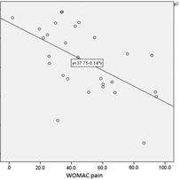
Correlation between gait analysis and clinical questionnaires in patients with spontaneous osteonecrosis of the knee
Abstract | Background: Spontaneous osteonecrosis of the knee is usually verified by magnetic resonance imaging accompanied by clinical questionnaires to assess the level of pain and functional limitation. There is a lack however, in an objective functional test that will reflect the functional severity of spontaneous osteonecrosis of the knee. The purpose of the current study was to examine the correlation between spatiotemporal gait parameters and clinical questionnaires in patients with spontaneous osteonecrosis of the knee.
Methods: 28 patients (16 females and 12 males) were included in the analysis. Patients had unilateral spontaneous osteonecrosis of the knee of the medial femoral condyle confirmed by magnetic resonance imaging. All patients performed a computerized spatiotemporal gait analysis and completed the Western Ontario and McMaster University Osteoarthritis Index and the Short-Form 36. Relationships between selected spatiotemporal gait measures and self-assessment questionnaires were assessed by Spearman non-parametric correlations.
Findings: Significant correlations were found between selected spatiotemporal gait parameters and clinical questionnaires (r ranged between 0.28 and 0.79). Single limb support was the gait measure with the strongest correlation to pain (r=0.58), function (r=0.56) and quality of life.
Interpretation: Spatiotemporal gait assessment for patients with spontaneous osteonecrosis of the knee correlates with the patient's level of pain and functional limitation there by adding objective information regarding the functional condition of these patients.
bottom of page


